
aschnell
New member
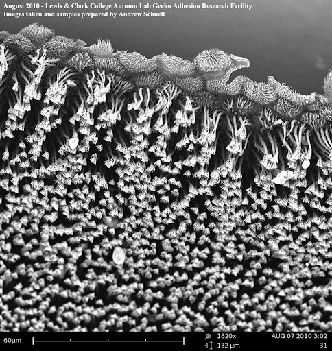
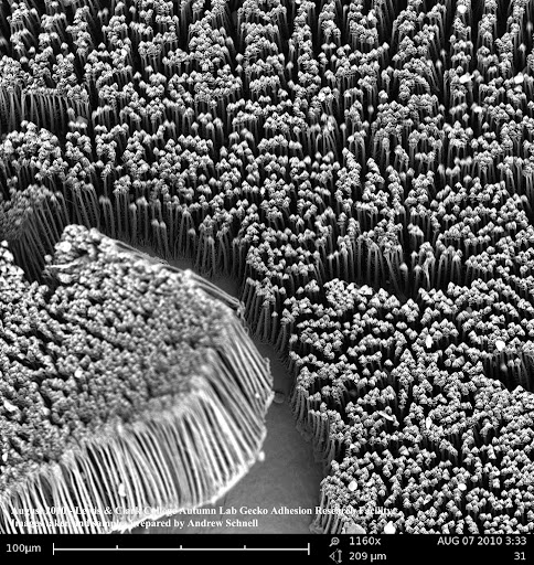
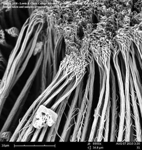
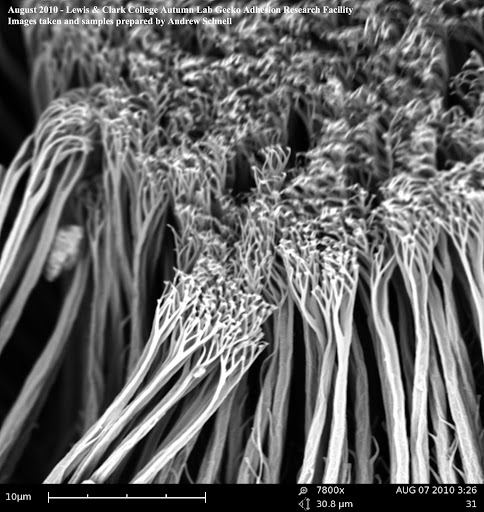
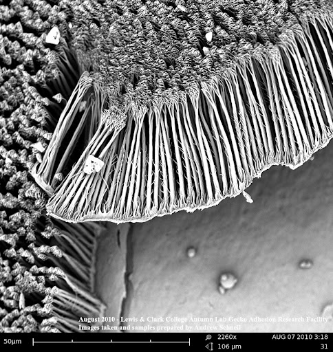
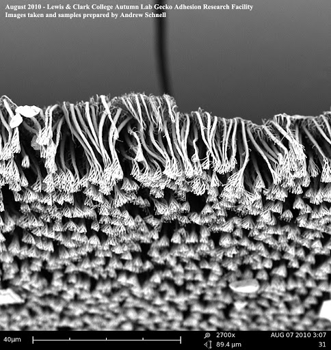
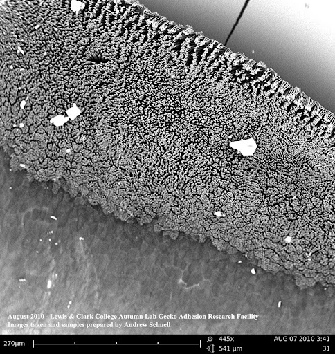
These are rare Scanning Electron Microscope Photographs of Rhacodactylus ciliatus setae (setae = the hairs on the bottom of gecko's feet). I prepared the samples and took these pictures using the scanning electron microscope here at Lewis & Clark College.
A scanning electron microscope works by bombarding a gold and platinum plated surface with a massive amount of electrons, and then reads how the electrons bounce off of the surface in order to create the image.
THESE PICTURES ARE COPYRIGHTED - Please don't save them to your computer
If you would like permission to have some of the images, just ask and I'd be happy to lend out the photos.
These images were difficult to take and are extremely valuable (not many SEM pictures of the setae of this species exist), so please think about what you're doing before you take them for yourself.
Anyway I thought it would be neat to share with you all the pictures of what is letting your gecko stick and what my work here at the lab is all about, enjoy!
") Although I would recommend not trying to squint too hard at your gecko's feet lol, you may go blind or crazy before you actually see the hairs
Although I would recommend not trying to squint too hard at your gecko's feet lol, you may go blind or crazy before you actually see the hairs